Painless and Radiation-Free Mammography: Just Lie Down and Relax
The importance of early detection in breast cancer cannot be overstated, as it significantly increases the chances of successful treatment. For many women, the thought of undergoing a mammogram can be daunting due to the discomfort and anxiety associated with the procedure. However, advancements in medical technology are paving the way for less invasive and more comfortable alternatives. One such promising development is the ultrasound computed tomography, a method that could revolutionize how we approach breast cancer screening.
In this article, we will delve into the intricacies of mammograms, explore the emerging technology of ultrasound-based imaging, and provide essential information for women considering these tests.
- Understanding how a conventional mammogram works
- Differences between conventional mammograms and ultrasound computed tomography
- Challenges associated with three-dimensional imaging
- Who should undergo mammography and when?
- Why mammograms are not typically performed before age 35
- Cost considerations of mammograms
- What to expect during a mammogram
- Understanding mammography results and cancer detection
- Are there any side effects associated with mammograms?
- Exploring advanced breast imaging technologies
Understanding how a conventional mammogram works
A mammogram is a crucial tool in the early detection of breast cancer. During this procedure, the breasts are placed between two plates that compress them, allowing a small amount of X-rays to pass through. These rays are captured by a detector that creates images of the breast tissue, which can be printed or digitally processed for further analysis.
This process, while effective, can be uncomfortable for many women due to the pressure applied during the compression. Additionally, the exposure to X-rays, even in minimal doses, raises concerns about potential long-term effects on health.
Differences between conventional mammograms and ultrasound computed tomography
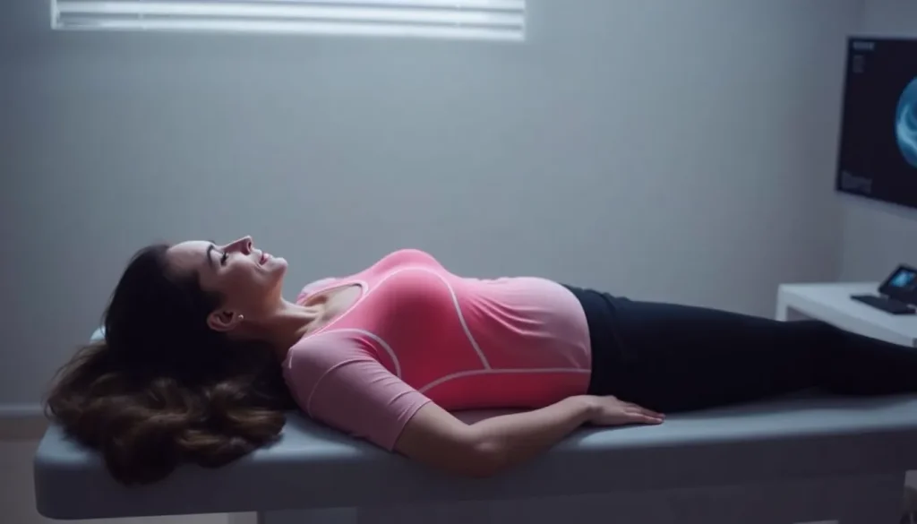
The ultrasound computed tomography represents a significant shift in breast imaging techniques. Instead of using X-rays, this method employs ultrasound waves, which are completely harmless. The patient lies face down on a specialized table with their breasts submerged in warm water, typically at a temperature of 36.5 ºC.
As the ultrasound waves pass through the breast tissue, they reflect off the various structures, creating detailed images of the internal anatomy. Unlike traditional mammography, this technique uses a conductive gel that facilitates the transmission of sound waves, allowing for a more thorough examination without discomfort.
- No radiation exposure
- Quick procedure, taking only about 3 minutes per breast
- More comfortable experience for the patient
- Potential for higher accuracy in detecting tumors
Challenges associated with three-dimensional imaging
One of the standout features of the ultrasound computed tomography is its ability to produce three-dimensional images. By utilizing advanced computational techniques, multiple two-dimensional images are captured and reconstructed to form a comprehensive 3D representation of the breast tissue.
In 2024, the Vall D'Hebron Hospital began conducting validation studies to evaluate the effectiveness of this new imaging technique in diagnosing breast cancer. Initial results indicate that it is quite effective, although further research is necessary to confirm its reliability as a mainstream diagnostic tool. For now, conventional mammograms remain a standard practice, but the future may hold less painful options for patients.
Who should undergo mammography and when?
Understanding when to start mammogram screenings is crucial for women’s health. Current guidelines recommend annual mammograms beginning at age 40 for most women, although those with a family history of breast cancer or other risk factors may need to start earlier.
- Women aged 40-44: Option to start annual screenings
- Women aged 45-54: Annual mammograms recommended
- Women aged 55 and older: Transition to biennial screenings or continue yearly based on preference
Why mammograms are not typically performed before age 35
Women under 35 are generally not advised to have mammograms unless they have specific risk factors. This is primarily due to the denser breast tissue in younger women, which can make it more challenging to detect abnormalities through mammography. Additionally, the radiation exposure, even at low doses, could potentially have adverse effects over time.
Cost considerations of mammograms
The price of a mammogram can vary widely depending on location, facility, and whether the patient has insurance. On average, a screening mammogram may range from $100 to $250. It's essential for women to check with their healthcare providers and insurance companies to understand coverage options and potential out-of-pocket costs.
What to expect during a mammogram
Preparation for a mammogram is straightforward, but knowing what to expect can help alleviate anxiety:
- Dress comfortably and avoid wearing deodorant or lotion on the day of the exam.
- Arrive early to complete any necessary paperwork.
- A technician will explain the procedure and guide you through the process.
- Your breasts will be positioned and compressed to capture the images.
- The exam usually takes less than 30 minutes, including preparation time.
After the procedure, results are typically available within a week, and your healthcare provider will discuss them with you.
Understanding mammography results and cancer detection
Interpreting mammography results can be complex. Radiologists look for various signs, including:
- Masses or lumps in the breast tissue
- Microcalcifications (tiny deposits of calcium)
- Changes in breast size or shape
If mammography reveals any suspicious findings, further tests such as ultrasound or biopsy may be necessary to confirm a diagnosis.
Are there any side effects associated with mammograms?
While mammograms are generally safe, some women may experience temporary discomfort due to breast compression. However, serious side effects are rare. It's crucial for women to communicate any concerns with their healthcare provider before the procedure.
Exploring advanced breast imaging technologies
As technology continues to evolve, newer imaging modalities such as digital breast tomosynthesis and MRI are becoming more prevalent in breast cancer screening. These methods may offer enhanced visualization of breast structures and improve early detection rates.
In particular, the introduction of ultrasound computed tomography brings hope for a painless, radiation-free alternative that could redefine breast cancer screening practices. Ongoing research will determine its potential as a reliable diagnostic tool.
For a visual representation of the advancements in imaging technology, consider watching this informative video:
The exploration of less invasive and more effective breast cancer screening methods is crucial in the fight against this pervasive disease. Women are encouraged to stay informed about their options and consult with healthcare providers to determine the best approach to their individual health needs.


Leave a Reply